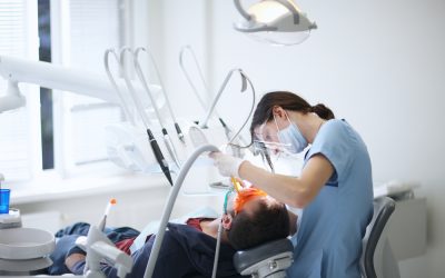
Cracked teeth are a common yet often undetected dental problem. They can lead to severe pain, infections, or even tooth loss if left untreated. Traditional methods for diagnosing tooth cracks, like visual inspection and X-rays, sometimes miss smaller or more hidden fractures. But thermal imaging dentistry is changing the way dentists approach this issue, offering a more accurate, non-invasive diagnostic tool to detect cracked teeth earlier.
In this post, we’ll explore how thermal imaging dentistry works, its benefits, and how it helps dentists pinpoint tooth cracks that might otherwise go unnoticed.
What Is Thermal Imaging Dentistry?
Thermal imaging dentistry is a modern diagnostic technique that uses infrared cameras to detect temperature variations in the teeth and surrounding tissues. The principle behind thermal imaging is straightforward: different surfaces absorb and emit heat differently. When there is a crack or damage to a tooth, the temperature in that area may vary slightly from the surrounding healthy tissue, which can be detected by the thermal camera.
This non-invasive technology is quick, safe, and doesn’t require the use of radiation, making it an excellent tool for both dentists and patients. It provides real-time, highly detailed images of the tooth’s surface, allowing for better detection of tooth cracks that might not be visible through standard examination methods.
How Thermal Imaging Helps Detect Cracked Teeth
One of the biggest challenges in detecting tooth cracks is that they often aren’t visible to the naked eye, especially if they are minor or located beneath the tooth’s surface. X-rays might miss tiny fractures, and visual inspections alone aren’t always sufficient, especially for cracks that occur in hard-to-see areas.
With thermal imaging dentistry, however, dentists can spot cracks with greater precision. When a tooth is cracked, it may have a different temperature profile compared to the surrounding, intact parts of the tooth. This is because cracks disrupt the tooth’s structure, affecting its heat retention and dissipation properties. The infrared camera picks up these subtle temperature differences, producing an image that clearly shows where cracks are located.
By using this technology, dentists can detect cracks that may not be immediately obvious, providing an early diagnosis that can prevent further damage and preserve the health of the tooth.
The Advantages of Thermal Imaging in Dentistry
There are several reasons why thermal imaging dentistry is becoming an essential part of dental diagnostics. Let’s take a closer look at some of the key benefits:
1. Non-invasive and Safe
Unlike X-rays, which use radiation, thermal imaging is completely safe and does not expose patients to any harmful radiation. It’s a completely non-invasive process, making it ideal for both adults and children. This makes it an appealing option for patients who are concerned about the risks of radiation exposure.
2. Early Detection of Tooth Cracks
The earlier a crack is detected, the easier it is to treat. Small fractures in a tooth can lead to bigger problems if ignored, such as infections or even tooth loss. By using thermal imaging dentistry, dentists can catch tooth cracks at their earliest stages, often before they are visible on X-rays or during a visual exam. Early detection means better treatment options and a higher chance of saving the tooth.
3. Accurate and Detailed
Thermal imaging provides highly detailed, real-time images of the tooth’s surface and surrounding tissues. This allows dentists to identify problems that are often missed with traditional diagnostic tools. Since thermal imaging can detect even minor temperature changes, it offers a higher level of accuracy in identifying tooth cracks.
4. Pain-Free and Quick
The procedure for thermal imaging is fast and painless. There’s no need for drilling, injections, or taking X-rays. Patients simply sit comfortably while the dentist uses the infrared camera to scan their teeth. The process typically takes just a few minutes, making it a convenient diagnostic tool for patients with busy schedules.
How Dentists Use Thermal Imaging to Diagnose Cracks
During a routine dental check-up, if a dentist suspects there may be tooth cracks that aren’t immediately visible, they may use thermal imaging dentistry to get a clearer picture of the problem. Here’s how the process works:
-
Preparation: No special preparation is needed for thermal imaging. The dentist will clean the area to ensure that no food particles or debris interfere with the results.
-
Scanning: The dentist uses a hand-held infrared camera to scan the tooth and surrounding areas. The camera detects temperature variations and sends the data to a computer, which generates a heat map image of the area.
-
Analysis: The dentist reviews the thermal image, looking for any abnormal temperature readings. Areas of the tooth with cracks will often show up as cooler or warmer than the surrounding tissue. This helps the dentist pinpoint the location and severity of the crack.
-
Further Action: Once cracks are identified, the dentist can determine the best course of action, whether it’s a filling, crown, or other dental procedure, depending on the severity of the damage.
Combining Thermal Imaging with Other Diagnostic Tools
While thermal imaging dentistry is a powerful tool for detecting tooth cracks, it’s not always used in isolation. Dentists often combine thermal imaging with other diagnostic tools, such as X-rays or visual examinations, for a more comprehensive assessment of oral health.
For instance, thermal imaging can identify cracks that may not be visible on an X-ray, while X-rays can provide more detailed information about the extent of the damage beneath the surface. By using a combination of diagnostic tools, dentists can develop a more complete treatment plan to address any issues they find.
The Future of Thermal Imaging Dentistry
As dental technology continues to evolve, thermal imaging dentistry is expected to become even more advanced and widely used. The technology may eventually be able to detect other dental issues, such as early signs of tooth decay, gum disease, or even oral cancer. As this diagnostic tool improves, it will help dentists provide better care and offer patients more effective treatment options.
In conclusion, thermal imaging dentistry is a revolutionary tool that allows dentists to detect tooth cracks more accurately and at an earlier stage than traditional methods. With its non-invasive nature, quick results, and ability to pinpoint hidden cracks, it’s becoming an invaluable asset in modern dental care. By catching issues early, dentists can help patients avoid more serious dental problems down the road, ultimately saving them time, money, and discomfort.
If you’re experiencing tooth pain or suspect a crack, be sure to ask your dentist about thermal imaging as part of your next check-up. It could be the key to preventing further damage and maintaining your smile for years to come.


